
Authors


Evelina London Children’s Hospital, part of Guy’s and St Thomas’ NHS Foundation Trust, has the largest fetal cardiac service in the UK and the longest established unit in Europe, overseeing 2,000 pregnancies a year for the diagnosis of congenital heart disease.
The hospital’s expert team of fetal cardiologists are at the forefront of pioneering technologies used for enhanced detection of congenital heart disease. The team use a range of novel techniques to produce high-definition images enhancing the management of even very complex heart problems both before and after birth.
Detecting structural heart problems with the heart
Congenital heart disease describes structural defects of the fetal heart which may have very serious consequences for the baby after birth. Congenital heart problems develop during the early stages of a mother’s pregnancy. If left undetected, abnormalities such as an underdeveloped valve, heart chamber, septal defect or abnormality of the major blood vessels may impact on wellbeing and even survival of the newborn.
In the middle-east the prevalence of congenital heart disease is reported to be higher compared to most other countries1. In the UAE for example, 1 out of every 100 babies is born with congenital heart disease, making heart problems the most common type of birth defect.
Dr Owen Miller, consultant in paediatric and fetal cardiology at Evelina London Children’s Hospital, explains, “It is important to precisely diagnose the type of congenital heart disease in the unborn baby during pregnancy. If the infant requires surgical intervention soon after delivery, we can prepare for surgery and achieve the best outcomes.”
A leading centre for three-dimensional fetal cardiac imaging
Evelina London Children’s Hospital works in partnership with research teams based at King’s College London and are at the forefront of cutting-edge fetal and paediatric cardiac diagnostic imaging research.
The teams have jointly developed a world-first method to conduct three-dimensional (3D) magnetic resonance imaging (MRI) scans which improves the ability to diagnose congenital heart disease in babies while still in the womb.
How the technology works
The hospital’s revolutionary MRI scanning technology uses unique motion-correction techniques to generate safe, comprehensive, high resolution 3D images of the fetal heart while still in the womb.
Dr David Lloyd, consultant in paediatric and fetal cardiology based at Evelina London Children’s Hospital, explains; “Using advanced methods like MRI is safe in pregnancy, but technically challenging as the fetal heart is so small and babies normally move a lot during the scan. This means standard MRI sequences cannot be used.”
“Our novel cardiac scanning method takes a series of two-dimensional MRI pictures of the fetal heart from different angles, then uses advanced computer technology which stitch the images together. This builds a high-resolution, three-dimensional image of the fetal heart and surrounding anatomy.”
Taking the technology one step further, these 3D images can then be converted into a physical representation of the cardiac anatomy, via 3D printing technology.
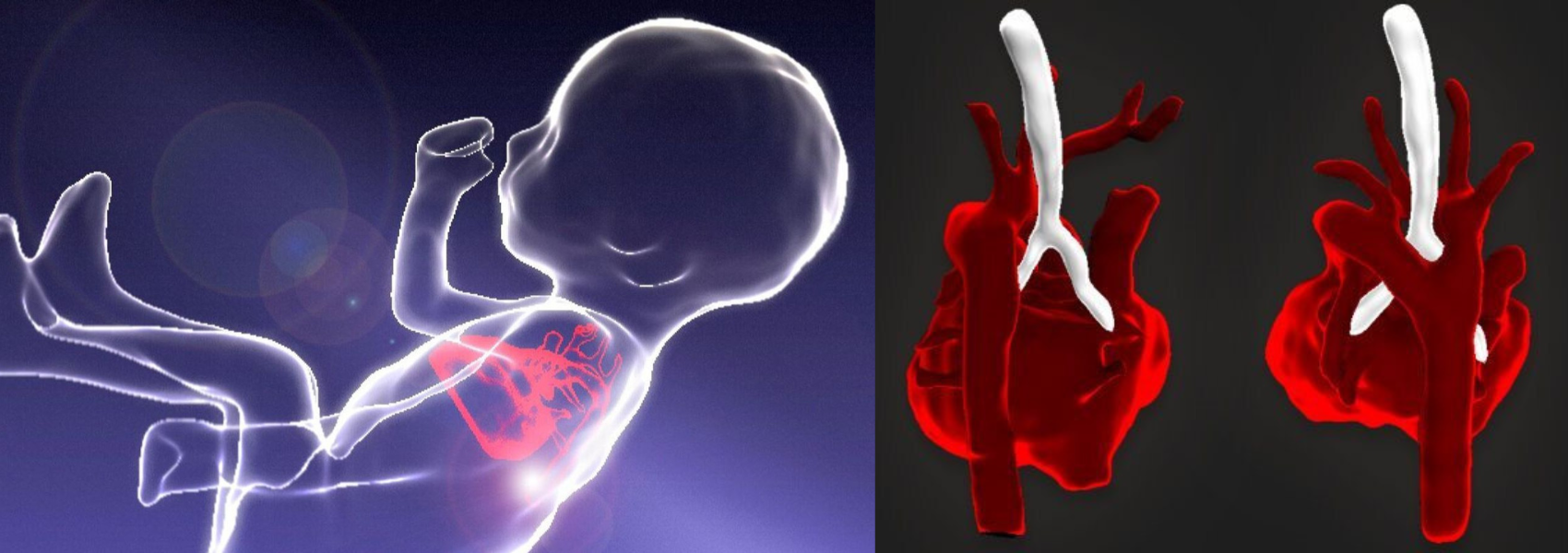
The technique transforms standard images into clear three-dimensional images (left). A 3D MRI scan of a fetal heart (right).
Improved care
Along with advanced ultrasound methods, these 3D MRI images can provide important insights into structural heart defects before birth, allowing the teams to plan treatment and improve our patients’ care before and after the child is born more accurately.
Dr David Lloyd explains, “This approach is now standard practice for the Evelina fetal cardiology team, who make a prenatal diagnosis of a cardiac abnormality in over 600 babies each year. It also improves the care of over 150 babies each year who need to deliver on site at St Thomas’s Hospital, London with known congenital heart disease.”
Looking to the future
The hospital’s team of scientists is now working to combine this 3D imaging with other advanced ultrasound and MRI techniques, to try to understand why some babies go on to develop more severe forms of congenital heart disease than others. Evelina London Children’s Hospital is the only UK centre to offer this ground-breaking approach. Contact our team to learn more.
Related content
-
Antenatal tests
As part of our leading private maternity service we offer a range of blood tests, swabs and screenings to support you during your pregnancy.
-
Cardiac imaging in children
Children's cardiac imaging helps diagnose heart diseases and defects. Diagnostics include echocardiography, cardiac MRI, CT and ECG.
-
Fetal cardiology
As one of the largest fetal cardiology units in Europe we manage heart problems in babies before birth.