
Content verified by

What is nuclear medicine?
Nuclear medicine is a scanning technique where small, safe amounts of radioactive materials, called tracers, are placed into your body, and using specialised scanners we take detailed images of organ function. The radioactive tracers are either injected, inhaled, or swallowed, and are developed specifically for the organ or system of your body being examined (such as your heart). The tracers are specifically chosen to mimic physiological processes so we can see internal organ function from outside with the gamma camera (SPECT scanner).
Nuclear medicine imaging primarily evaluates how organs and tissues function (their physiology), and they are often combined with a CT scan, which provides 3D cross-sectional anatomical detail. This fusion of functional and anatomical images allows us to assess both how tissues and organs behave (whether they are working as they should) and how they look (whether there are any structural changes or damage).
Our consultant nuclear medicine physicians at Royal Brompton and Harefield hospitals are world-leading experts in cardiopulmonary imaging and other diagnostic nuclear medicine scans. We can provide a fast and accurate diagnosis for specific conditions in various organs/systems in both adults and children, using our 2 state-of-the-art nuclear medicine scanners.
Along with our expert doctors, we are a multidisciplinary team consisting of cardiac nurse specialists, highly skilled technologists, experienced medical physicists, courteous administrative staff, and supportive healthcare assistants.
Experts in nuclear medicine
We offer:
- fast access to nuclear medicine scans, in as little as 24 hours
- expert review by world-leading nuclear medicine consultants for the most accurate diagnosis
- clear and reliable scan results within 1 week

Why would you need a nuclear medicine scan?
Nuclear medicine scans are used to diagnose conditions (sometimes even in their earliest stages), to monitor how well a treatment is working, or to help plan future treatment. The images produced provide unique information that is often not available from other imaging techniques, showing not only the structure of your body but also important functional changes.
Nuclear medicine scans can be used for assessing how different organs or systems work
- heart – checking blood supply to the heart muscle, the effects of a heart attack, identifying which parts of the heart muscle may benefit from procedures like coronary artery bypass surgery or angioplasty, as well as to assess the fitness of your heart before surgery
- lung – to investigate acute or chronic blood clots in the lungs (pulmonary embolism) and comparing that with the aeration of your lungs. It can also be used to assess the effect of chronic airways disease on lung function and deciding the type of corrective surgery, as well as to evaluate breathlessness related to long COVID
- bones – assessing subtle fractures, bone secondaries and evaluating joint prosthesis and their complications such as infection/inflammation
- gastrointestinal system – such as assessing gastro-oesophageal reflux or delayed digestion
- infections – to pinpoint the precise location of infection for more effective treatment with a white cell scan
- kidneys – to check if they are working as they should and if there are any scars on the kidneys
- lymphatic system – to assess lymphatic drainage or identify blockages
- brain – for evaluating Parkinson’s disease
- thyroid – to assess the functional ability in various thyroid-related diseases
- specific types of cancer – diagnosis of neuroendocrine tumours, such as phaeochromocytoma
If you have been referred for a nuclear medicine scan, our team can arrange a private appointment at a time that suits you. Contact us.
Our advanced nuclear medicine scanners
We use the VERITON-CT and DSPECT scanners for our patients at Royal Brompton Hospital. With their advanced detectors, they enable faster scans, lower levels of radiation for improved safety and better image quality. Both scanners are among the most advanced in the field and are the only examples of their model in London, with limited availability in other parts of the UK.
With this innovative technology in our hands, we can provide precise structural and functional 3D scans of the heart, lungs and other areas of the body. This helps our specialists to understand disease progression (for conditions like pulmonary embolism) and to plan the best treatment (such as identifying which part of the heart will benefit most from angioplasty).
VERITON-CT
Our state-of-the-art SPECT-CT scanner at Royal Brompton Hospital, called the VERITON-CT is the first of its kind in London and is the most advanced nuclear medicine scanner available.
It combines SPECT (single-photon emission computed tomography) with CT imaging in a single imaging session. By combining the imaging technologies, it enables us to see the structure and function of the heart and lungs, gathering comprehensive information about a patient’s health in one scan.
With 12 highly sensitive detectors arranged in a 360-degree layout around the body, the VERITON-CT produces fast, high-quality 3D images, reducing the need for additional diagnostic tests. Sometimes we can do two tests at the same time (ventilation and perfusion scan) reducing the scan time and improving patient comfort.

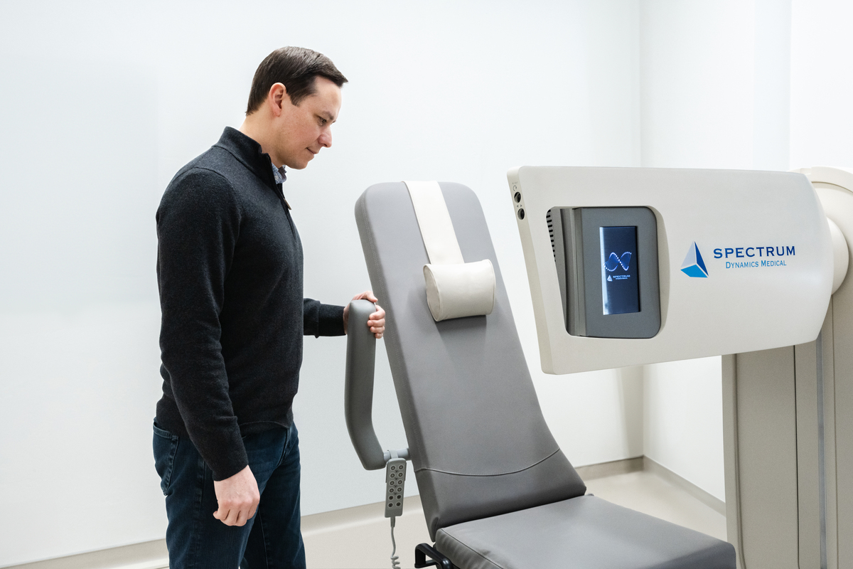
DSPECT
The DSPECT is an advanced dedicated cardiac scanner with detectors which only circle the left and top of your chest. This allows us to acquire a high-resolution image without enclosing your head and can be completed while you are seated.
With its innovative design, we can scan the hearts of any patient, regardless of your level of mobility or BMI – we can scan patients up to a weight of 175kg. Its open design is also beneficial for patients with claustrophobia. Moreover, patients with metallic implants and impaired kidney function can have all types of nuclear scans. Overall, the DPECT helps create a more comfortable experience for patients undergoing a nuclear medicine scan.
What are the types of nuclear medicine scans?
We offer a wide range of nuclear medicine scans, using different tracers that either bind to specific tissues or organs, or circulate through various systems of the body. Please find a list below of the common nuclear medicine scans we provide.
A myocardial perfusion scan determines the blood supply to your heart by using a small amount of a radioactive tracer called tetrofosmin/sestamibi, which is injected into a vein in your arm. It allows our consultants to view your heart under stress and rest, to assess whether there is any occlusion (blockage) or shortage of blood supply to the heart muscle.
There are several benefits of having this type of scan at Royal Brompton Hospital, including:
- fast scan times – typically between 2 and 10 minutes per scan
- the option to have scans performed while seated
- an open scanner design, which is particularly helpful for patients with claustrophobia
- a highly specialised team with world-leading expertise in heart care
A VQ scan enables us to assess the aeration (ventilation) and blood flow (perfusion) to your lungs. It involves inhaling a very small amount of radioactive gas called krypton and injecting a very small amount of tracer called MAA into a vein in your arm.
The krypton gas allows us to see the airways in your lungs and the MAA tracer looks at blood flow to your lungs. The comparison of these two assessments allows us to evaluate the presence of a pulmonary embolism (blood clot in the lungs), as well as other lung diseases.
At Royal Brompton Hospital, these scans are performed with the highest image quality, at around half the usual radiation dose, and in roughly a third of the time compared with other sites.
A cardiac amyloidosis imaging scan maps the amount of an abnormal protein called amyloid in your heart tissue. Amyloid can cause the heart muscle to stiffen, making it harder for the heart to pump blood around the body.
During the scan, a small amount of a tracer called 99mTc-DPD is injected into a vein in your arm. The tracer collects in certain areas of the heart muscle, allowing us to identify the presence of amyloid.
The results help our consultants confirm conclusively whether the disease is present and plan the most appropriate treatment for you.
Bone scans can be performed for various indications, including for cancer metastasis (spread of cancer) and bone fractures, infections or inflammation.
We inject a small amount of tracer called HDP that allows us to visualise bone metastasis, infection and inflammation. At Royal Brompton Hospital, the scans are completed as 3D images to visualise the conditions in more detail than 2D offered at other centres
A DaTSCAN helps us to visualise the dopamine transporters in the brain, which are correlated with the presence of Parkinson’s disease. We inject a tracer called ioflupane into a vein in your arm, which binds to dopamine transporters in the brain. If the scan shows abnormal pattern in the brain, it indicates the presence of the condition.
A DMSA kidney scan is used to detect any damaged/scarred areas of the kidneys, for example because of repeated urinary tract infections. It also helps us see whether your kidneys are working equally well. We inject a small amount of a tracer called DMSA into your arm which is absorbed by the kidneys and shows up with a scan.
A renal MAG3 scan allows us to see how well your kidneys are working, by using a tracer called MAG3 that we will inject into a vein in your arm. The scan helps us visualise how blood flows to your kidneys, how urine is draining from your kidneys and whether there are any blockages in your urinary system. It also helps assess whether you may require a kidney transplant.
A gastric emptying scan determines how quickly your stomach empties after a meal, to understand whether you have a condition called gastroparesis – delayed digestion.
We will ask you to eat a meal that is mixed with a small amount of tracer before we will take images every 15 minutes for 2 hours. With these images, we can track how your food moves through your stomach and intestines over time.
A gastro-oesophageal reflux scan, also known as a ‘milk scan’, assesses whether there is an abnormal return of stomach contents back into your oesophagus (food pipe). It is also another way of measuring how fast your stomach empties.
We will ask you to drink milk/juice/ liquid with a small amount of tracer in it before the test. During the test, you will lie on our scanner bed for 1 hour while we take multiple images.
An Iodine-123 MIBG scan helps us diagnose and assess certain types of cancers that start in hormone-producing neuroendocrine cells, including neuroendocrine cancers, neuroblastomas, medullary thyroid carcinomas (a type of thyroid cancer) and phaeochromocytomas (a type of adrenal gland cancer).
In addition to detecting cancer, an Iodine-123 MIBG scan can be used to check the neuronal function of the heart. This is particularly useful for patients with heart failure, to help our heart specialists understand the underlying causes of the condition and tailor treatment accordingly.
White blood cells are an important part of your immune system, defending you from infection. They collect in areas where your tissues have become infected or inflamed. If your doctor suspects that you have an internal area of infection or inflammation and want to determine the precise location to provide the best treatment, they may refer you for a labelled white cell scan.
As part of the scan, we will take a small blood sample (50ml) and extract your white blood cells from it. We will mix these white blood cells with a small amount of tracer that binds to them (a ‘label’) before injecting these cells back into you via a vein in your arm.
A lymphoscintigraphy is a type of nuclear imaging test that is used to create detailed images of your lymphatic system, as well as to locate your lymph nodes. The lymphatic system helps protect you from infection and keeps a healthy balance of fluid throughout your body.
This test helps us understand how well your lymphatic system is working and particularly if there is blockage, causing a swelling (a condition called lymphadenopathy).
We will inject a small amount of tracer into a vein in your arm that circulates throughout your lymphatic system before taking images.
A thyroid scan shows how well your thyroid gland is functioning. We inject a small amount of tracer into a vein in your arm, which is then absorbed by your thyroid gland. By measuring how much tracer your thyroid takes up, we can assess whether you have hypothyroidism (underactive) or hyperthyroidism (overactive).
This test is also used to help diagnose Graves’ disease (an autoimmune condition in which your immune system attacks the thyroid gland), as well as other thyroid disorders.
Sentinel node localisation is used for breast cancer patients shortly before surgery. A sentinel node is the first lymph node that breast cancer is likely to spread to – it’s usually located in your armpit. A small amount of a tracer is injected beneath your nipple and this slowly travels to your sentinel node.
During surgery, surgeons will use a device called a gamma probe to detect the tracer and accurately locate and remove the sentinel node. Once removed, this will be tested for the presence of cancer cells. This helps our consultants understand whether your cancer has spread and its stage.
A DEXA scan (also known as a DXA BD scan) is a quick, highly accurate and non-invasive imaging test that measures your bone density to diagnose or assess the risk of osteoporosis. It uses low-dose X-ray beams to scan your body, making it much safer than standard X-rays.
Osteoporosis is a condition that causes your bones to become weaker and more fragile, making them more likely to break or fracture. There are several risk factors, including:
- age: as we age, the natural breakdown of bone cells outpaces the creation of new ones, with people over 50 at greater risk of osteoporosis
- sex: women lose bone mass more rapidly than men as they age, particularly after menopause due to reduced levels of the hormone oestrogen
- lifestyle: a sedentary lifestyle (not exercising enough), smoking and excess alcohol consumption can all increase your risk
- medications and health conditions: medications like steroids can increase your risk, as can conditions like rheumatoid arthritis and hyperthyroidism
We use the GE Lunar IDXA scanner at Royal Brompton Hospital, which uses state-of-the-art fan beam technology. This enables us to complete scans much faster with images that are of higher quality for greater accuracy. Overall, with a quicker scan, it provides you with a more comfortable experience.
Preparing for a nuclear medicine scan
Each nuclear medicine scan is unique to the tissue or organ being imaged. This means that there isn’t a standard way of preparing for your scan and that you will be provided specific instructions beforehand that you will need to read through. However, below is some general guidance that is common to all nuclear medicine scans.
Before your scan, you may be asked to avoid certain foods (such as those containing caffeine) or to fast for a period. In some cases, you might also need to pause certain prescribed medications, as they can affect how the organ or tissue appears on the scan. We will give you clear instructions in advance, so you know exactly what to do. Please don’t hesitate to get in touch on 020 7351 8666 if you wish to speak with us before your appointment.
On the day of your test, please try not to bring valuables or jewellery. These can get lost easily, and jewellery can sometimes interfere with the scan. You may also be asked to change into a hospital gown to make it easier for us to get the images.
Please arrive at least 15 minutes before your appointment so our team has time to prepare you, and to help you feel relaxed before your scan.
We will first start by taking your medical history and confirming your details. An explanation of the scan will be followed by us providing the tracer needed for imaging. This can be given intravenously (such as through a vein in your arm), inhaled or swallowed, depending on the type of test and specific are of your body being examined.
There is minimal amount of radiation in the tracer, so nuclear medicine scans are very safe, with no side effects and generally no allergic reactions. Your consultant or doctor will only recommend them in cases where they need important information that can’t be provided with other types of tests.
The duration of a nuclear medicine scan is different for every type, but they will usually be less than 1 hour, with some requiring more time and repeat visits to our hospitals. You may also need to wait for a short time between receiving the tracer into your body and getting the scan. If this is the case, you won’t need to stay and can leave, with our technologists informing you of when to return for your scan.
During the scan, we will ask you to lie on your back and stay as still as possible. Images of your body will be captured using a nuclear medicine scanner to record the distribution of the tracer in your body. This is often combined with a CT scan.
You will be allowed to go home after your scan and will usually be able to resume your normal activities and diet immediately. However, as tracers emit small levels of radiation, you may be given instructions of what to do after your scan as a precaution. These depend on the type of scan performed and are often related to breastfeeding and keeping distance from small children and pregnant women. Rest assured our technologists will inform you of what you need to do on the day.
Getting your nuclear medicine scan results
After your nuclear medicine scan is reviewed by one of our specialist consultant nuclear physicians, your results will be sent to your referring consultant who will discuss them with you.
You won’t get your results immediately after your scan. It can typically take between 1 and 3 days before they are ready – you can confirm the waiting time with your referring consultant.
If you are diagnosed with a condition or your treatment needs to be changed following the results of your scan, your referring consultant will talk you through the next steps.
Nuclear medicine scan pricing
The cost of a nuclear medicine scan will vary depending on the type of scan, complexity, and individual requirements. We offer transparent pricing and will always confirm costs with you before your appointment.
| Type | Price from |
|---|---|
| Bone scan for assessment of bone infection/inflammation | £1,950 |
| Bone scan for assessment of cancer metastasis | £1,828 |
| Cardiac amyloidosis imaging (99mTc-DPD scan) | £1,928 |
| DaTSCAN for Parkinson’s disease assessment | £2,110 |
| Gastric emptying scan | £357 |
| Gastro-oesophageal reflux (milk) scan | £357 |
| Iodine-123 MIBG scan for cardiac innervation | £1,361 |
| Iodine-123 MIBG scan for neuroendocrine cancers | £1,361 |
| Labelled white cell scan | £2,240 |
| Lung ventilation and perfusion (VQ scan) | £2,162 |
| Lymphoscintigraphy | £357 |
| Myocardial perfusion scan | £1,672 |
| Renal MAG3 scan for assessment of kidney function | £418 |
| Renal DMSA scan for assessment of kidney disease | £418 |
| Sentinel node localisation | £357 |
| Thyroid scan | £296 |
Reviewed regularly to reflect clinical best practice
Last reviewed: 14 November 2025
Location
Meet our team of nuclear medicine specialists
At Royal Brompton Hospital, our consultant nuclear medicine physicians are internationally recognised for their expertise in clinical work, research and teaching. Using our advanced VERITON-CT and DPSECT scanners, we deliver fast, accurate nuclear medicine scans for adults and children, helping diagnose a wide range of conditions with precision and care.