
Content verified by


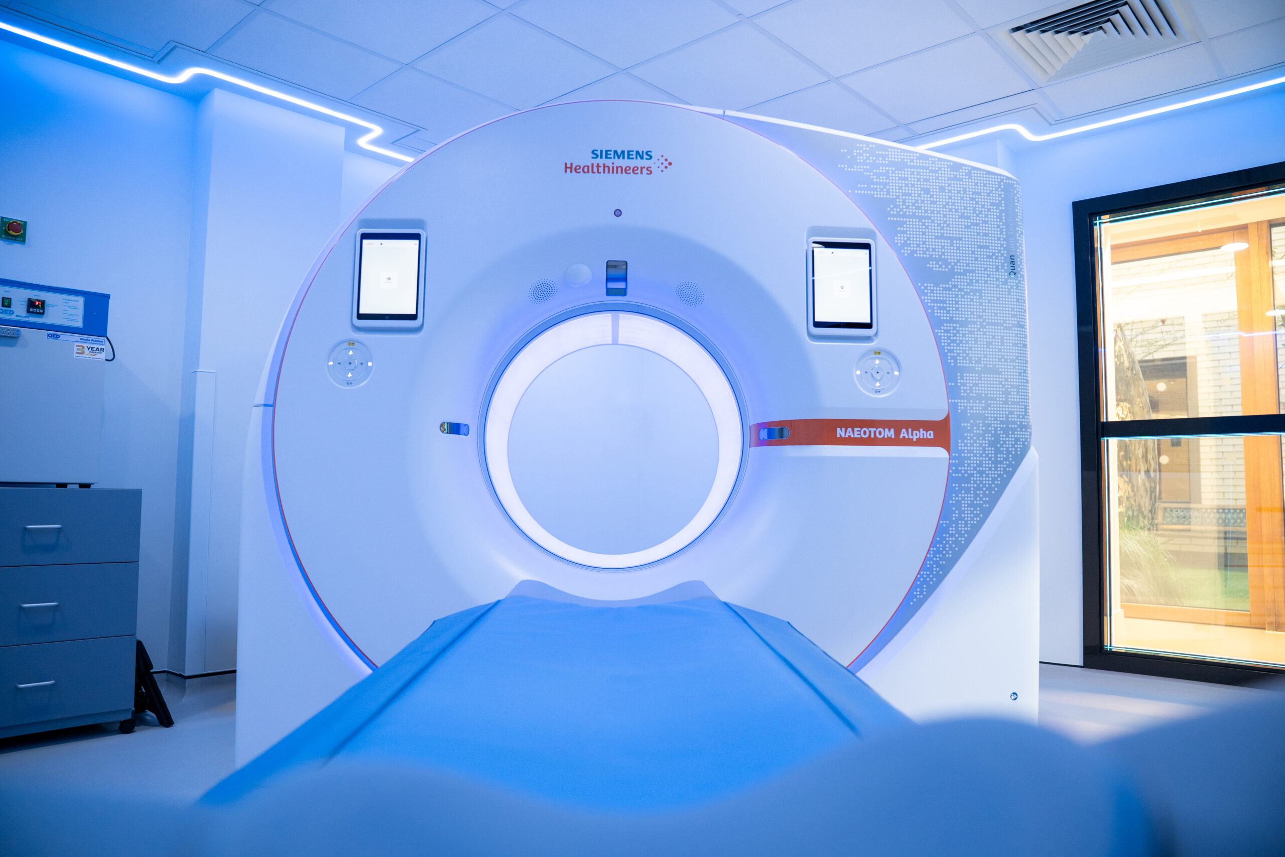
NAEOTOM Alpha CT scanner at Wimpole Street Consulting Rooms and Diagnostic Centre
The NAEOTOM Alpha is the world’s first commercially available photon-counting CT scanner (PCCT). Rather than converting X-ray photons into light and measuring the light, PCCT converts X-rays directly into electrical signals, allowing them to be individually counted. This can produce a never–before-seen level of image detail and has the potential to lower radiation doses required for imaging at a quality above and beyond any other CT technology available in the private sector today. Learn more about the NAEOTOM Alpha from Professor Ed Nicol, Dr Thomas Semple, and Dr Jonathan Weir–McCall, consultant cardiologists and radiologists at Royal Brompton Hospital.
Traditional CT scanners versus the NAEOTOM Alpha
Traditional CT scanners use X-ray systems and advanced computing to produce detailed 3D images of structures inside the body. This technology works well for static structures and organs where motion can be controlled (for example lung imaging with a short held-breath). However, for organs that move when the patient is at rest – such as the heart – a traditional CT scan results in motion-induced blurring of images.
For cardiac CT scans, patients are often given medication (beta blockers) to slow down the heart for clearer imaging of the coronary arteries. However, for fast or irregular heart rates, imaging with traditional CT scanners may still be challenging.
Royal Brompton Hospital has been at the forefront of cardiovascular CT for decades, most recently by using Siemens dual source CT machines. These scanners are, by design, twice as fast as conventional CT systems, making them better suited for cardiac CT imaging. The NAEOTOM Alpha CT scanner is Siemens’ most advanced dual source CT scanner and adds additional capabilities above and beyond that possible with traditional detector technology.
We’re proud to share that Wimpole Street Consulting Rooms and Diagnostic Centre is now home to a NAEOTOM Alpha CT scanner – the first of its kind in private practice in the country – offering cutting edge diagnostics to our patients.
Benefits of the NAEOTOM Alpha
“The NAEOTOM Alpha will allow us to freeze heart motion, producing exquisite diagnostic quality images of the coronary arteries and other heart structures in a fraction of a second, at the lowest possible radiation dose, even when the heart rate is high or irregular, for example in atrial fibrillation,” explains Professor Nicol.
Further to this, the scanner’s revolutionary detector allows our specialists to see inside coronary stents as small as 2mm in diameter (well below the limits of resolution for traditional CT scanners), as well as reducing artefact from metal objects like pacemakers and the larger stents found around TAVI valves.
With its precise imaging, our specialists can better characterise the composition and severity of coronary artery stenosis caused by atheromatous plaque, reducing the need for invasive coronary catheter examinations or further investigation. This provides improved accuracy for characterisation of the disease present in coronary arteries, to help our consultants develop the best-informed treatment strategy for patients.
While the NAEOTOM Alpha CT scanner can be used to scan any part of the body, it is revolutionary for cardiac patients, particularly those who:
- have had previous coronary intervention, such as a stent
- have pacemakers or other metal implants
- require repeat CT imaging
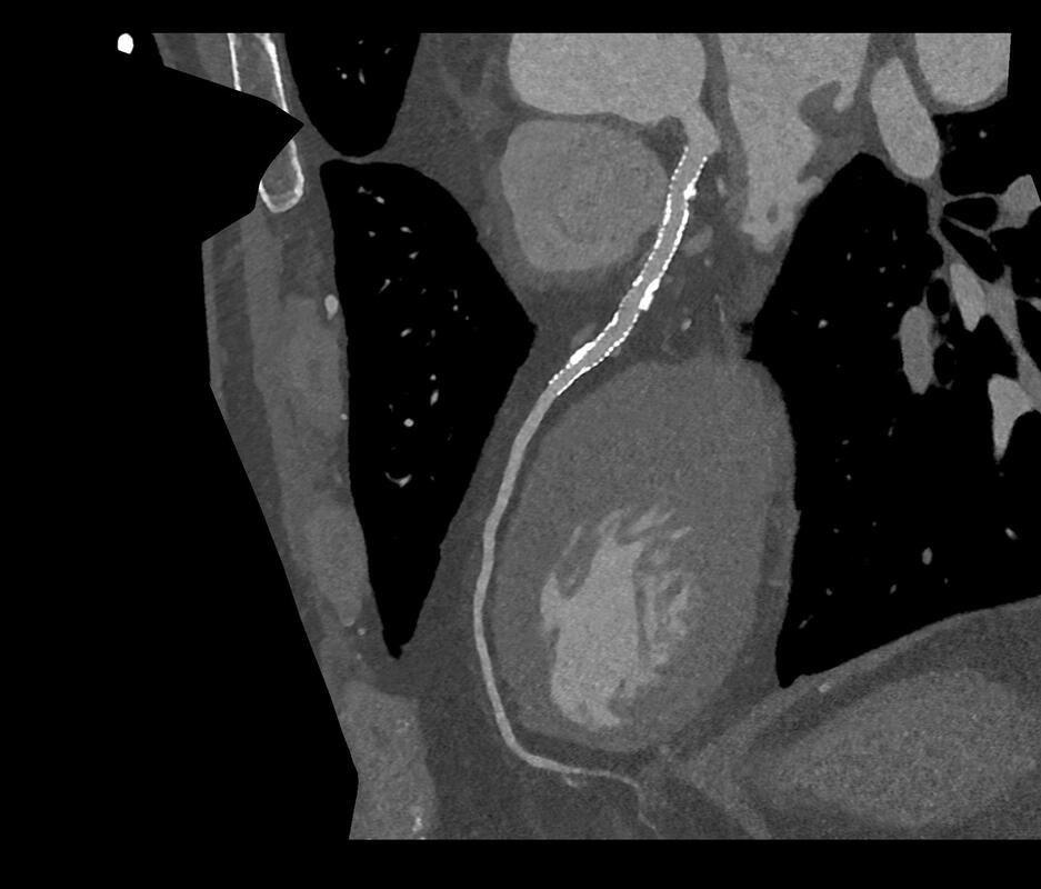
In-stent evaluation becomes feasible in clinical routine, thanks to the unique combination of resolution, spectral capabilities, and high temporal resolution of the NAEOTOM Alpha CT scanner. Image courtesy of Center Cardio-Thoracique de Monaco, Monaco via Siemens Healthineers.
Enhanced safety and precision
One of the key benefits of the NAEOTOM Alpha CT scanner is in balancing image quality with radiation exposure. “This machine has the potential to allow clinical imaging at the lowest radiation dose of any commercially available scanner ever made,” explains Dr Semple. “For patients with cardiovascular risk factors such as high blood pressure or a family history of coronary artery disease, we now have the capability to offer a scan with high enough resolution to exclude any coronary disease at a very low dose.”
Centres outside of the UK have achieved up to a 45% reduction in radiation dose when compared to traditional CT scanners, with no effect on image quality, across all body areas and up to 50% reduction in dose for lung imaging, without any loss of image quality.
Additionally, we are very proud of our active imaging optimisation program, with imaging and physics specialists meeting regularly to ensure that every scan we perform meets our requirements for optimal image quality at the right dose. Dose monitoring software allows real-time capturing of trends over time and helps ensure our service maintains its very high standards. This should be particularly reassuring for those requiring multiple follow up scans.
Quality images without compromising speed
In terms of image quality, the NAEOTOM Alpha provides slices as small as 0.2mm (compared to 0.5mm for traditional CT scanners), even for patients with a high BMI. This resolution is the highest available for any CT scanner currently, which greatly increases diagnostic confidence. It is this very high resolution that enables our specialists to assess for blockages even within small coronary stents, a task that is often not possible via traditional CT technology.
Some of the advanced applications offered by the NAEOTOM Alpha can be provided by dual energy, or older spectral CT scanners, but only at a very slow speed. While this may be acceptable for other organs, the NAEOTOM Alpha is the only scanner to allow these techniques to be applied at the speed required for high-end cardiovascular imaging, with a scan speed of up to 74cm per second, in line with the fastest traditional CT scanners, but with the significantly increased image quality, decreased radiation dose and advanced imaging capabilities.
Versatility across medical specialties
While the scanner will initially mainly benefit cardiac patients at Wimpole Street – due to the significant improvements from standard heart scans offered currently – it can also be used for investigations across a range of medical specialties. “The NAEOTOM Alpha is a huge leap forward in terms of cardiac diagnostics,” says Dr Weir-McCall, “however, it can also be applied to other clinical indications to plan interventions.”
Respiratory patients will also benefit from the clearer depiction of airways and fine detail of lung structure, increasing diagnostic confidence. Young patients requiring frequent imaging (such as those with cystic fibrosis or other chronic lung diseases) will also benefit from the low radiation dose capability.
When to refer to us
The NAEOTOM Alpha CT scanner is suitable for any adult patient requiring a CT scan, but its capabilities are particularly revolutionary in the care of patients who have previously had coronary intervention or have established coronary artery disease. Patients who require assessment of coronary stents, for example to detect restenosis, would particularly benefit from the NAEOTOM Alpha, as they may be unsuitable for traditional CT scans.
Patients referred to us for private diagnostics at Wimpole Street Consulting Rooms and Diagnostic Centre can be seen quickly, with a personalised care plan developed if they require further treatment.
Get in touch
To find out more about the NAEOTOM Alpha CT scanner and our imaging expertise, please contact our customer services team. Call +44 (0)20 3131 5130 or email privatepatients@rbht.nhs.uk
Related services
-
CT scan
A CT uses X-rays and a computer to produce images of many structures inside the body.
-
Diagnostic tests for heart conditions
We offer a range of diagnostic tests at our state-of-the-art facilities.
-
Wimpole Street Consulting Rooms and Diagnostic Centre
Our state-of-the-art outpatient and diagnostics centre brings experts from across our hospitals to one convenient central London location