
Content verified by

What is retinal detachment?
Your retina is a thin layer of light-sensitive tissue at the back of your eye. In retinal detachment, the retina pulls away from its original position, causing it to lose its blood supply.
Retinal detachment is a serious eye condition that requires urgent retinal detachment surgery (vitrectomy) to prevent vision loss.
At St Thomas’ Hospital, our ophthalmology team can promptly diagnose and treat retinal detachment to protect your eye health and vision.
Experts in retinal detachment
Our ophthalmology specialists at St Thomas’ Hospital offer:
- short notice appointments
- fast and accurate diagnosis
- fast access to treatment
Who is at risk of retinal detachment?
Some people are at a higher risk of retinal detachment than others.
Risk factors that may increase your likelihood of retinal detachment include:
- myopia or short sightedness
- severe eye injury
- previous eye surgery
- a family history of the condition
- other eye diseases including diabetic retinopathy
- previous retinal detachment in one of your eyes
If you are worried about your risk of developing retinal detachment, speak to our team of highly experienced ophthalmologists by completing our online enquiry form today.
Types of retinal detachment
There are 3 types of ways that your retina can detach from the back of your eye:
- rhegmatogenous retinal detachment
- tractional retinal detachment
- exudative retinal detachment
A tear or hole in your retina, causing fluid to pass through and build up, causes rhegmatogenous retinal detachment. Eventually, your retina pulls away from its supporting tissues. This is the most common cause of retinal detachment.
Tractional is where scar tissue grows on the retina’s surface, causing it to pull away from your eye. This type is more common if you have unmanaged diabetes, although it’s a less common type of retinal detachment.
With exudative retinal detachment, fluid builds up without a tear in your retina. Age-related macular degeneration (an eye condition affecting central vision), eye injuries, tumours and inflammation are the typical causes of this type of retinal detachment.
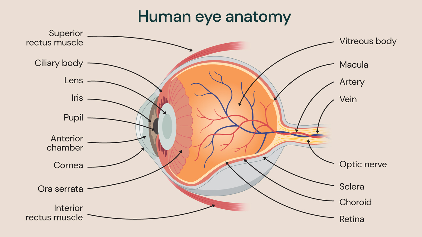
Illustration showing the anatomy of the human eye.
Retinal detachment symptoms
The symptoms of retinal detachment can happen suddenly, especially if the detachment is severe.
Symptoms of retinal detachment include:
- floaters in your vision (seeing flecks, dark spots or wiggly lines floating)
- darkening of your peripheral (side, top or bottom) vision
- shadows covering parts of your vision
- flashes of light (especially when moving the eyes and in the dark)
It’s vital to watch for any changes in your vision, as retinal detachment can cause permanent damage to your sight.
If you are experiencing retinal detachment symptoms, book an appointment with one of our specialists who can help with a diagnosis.
What causes retinal detachment?
Retinal detachment is generally caused by changes to the jelly substance in your eye, which is called the ‘vitreous’.
These changes can happen as you get older, leading to a condition called posterior vitreous detachment (PVD). It’s PVD that can lead to retinal detachment due to the creation of retinal tears and holes after separation of the vitreous. There is no known way to prevent PVD.
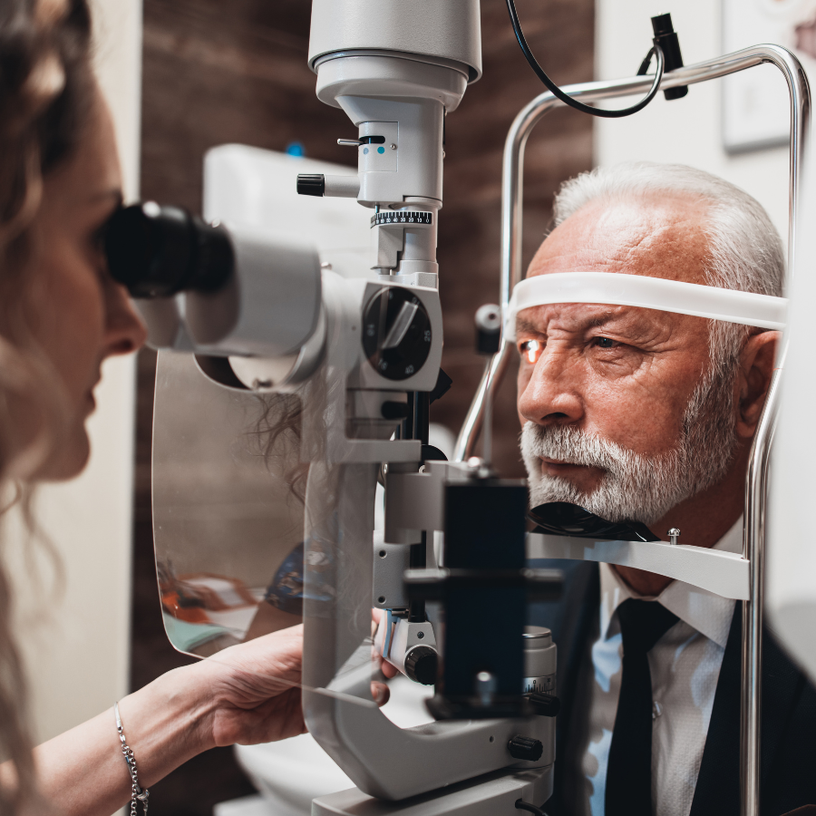
How is retinal detachment diagnosed?
Ophthalmologists can diagnose retinal detachment in various ways. The main method used involves your ophthalmologist examining your retina using a bright light with a lens attached, allowing them to see the back of your eye.
Your ophthalmologist will use special eye drops to dilate (widen) your pupils (the black part of your eye) to see your retina more clearly and check for holes or tears.
If they cannot see into the back of your eye, they may arrange for ultrasound imaging tests. They will typically examine both eyes even if you only have symptoms in one.
Retinal detachment treatment
Retinal detachment needs treatment as soon as possible to reduce the risk of permanent vision loss and damage.
Retinal detachment surgery (vitrectomy) is the primary treatment for retinal detachment.
In a vitrectomy, the ophthalmologist removes the vitreous, leaving a space and inserting a gas or silicone oil bubble. Depending on the type of gas used, it will absorb spontaneously by itself between 10 days and 8 weeks. Silicone oil will require further surgery to remove the oil from the eye, and can be carried out as a day surgery.
The gas bubble helps to push the torn part of your retina against the wall of your eye, preventing fluid from flowing through the holes. The fluid build-up in your eye will be absorbed into your body and your retina should reattach.
We can perform vitrectomies under either general (where you sleep through the procedure) or local (where only your eye is numbed) anaesthetic. If you are under local anaesthetic, you will be awake but unable to feel any pain.
You can usually go home on the same day as your treatment. Due to the gas bubble in your eye, your distance vision will be poor, so you cannot drive yourself home. Your vision should improve once your body has absorbed the gas.
Risks of vitrectomy
All surgeries carry potential risks. However, our committed team works hard to minimise them and provide you with safe treatment.
The possible risks of a vitrectomy include:
- failure to repair your retina
- infection
- increased eye pressure, elevating your glaucoma risk
- elevated risk of cataracts
- changes to your vision (blurring, distortion)
Your consultant will explain the potential risks before surgery so you are fully informed. Although vitrectomies have some risks, our surgeons’ wealth of knowledge and experience helps reduce them and provide you with effective results.
Book an appointment with Guy’s and St Thomas’ Specialist Care
If you are concerned about retinal detachment, please seek immediate medical attention. Retinal detachment is a serious condition that needs urgent treatment to save your vision.
Book a consultation with our ophthalmology team today and discover how we can support your vision and eye health.
Reviewed regularly to reflect clinical best practice
Last reviewed: 09 March 2026
Locations
Our ophthalmology service is located at:
Discover our ophthalmology specialists
Meet our team of world-class ophthalmologists. From vision assessments to helping with conditions like cataracts or glaucoma, access private eye care in London.